It is a common condition in young children and involves the deviation of one or more toes. In many cases, it corrects spontaneously, although some patients may require the use of orthotics or even surgery.
It is one of the most common anomalies of the fingers and involves the duplication of a finger, mainly the big or little toe. The treatment is surgical.
It is the partial or complete fusion of two or more toes. Most cases do not require treatment as they do not cause any limitation in mobility.
The front half of the foot is deviated inward. It is a condition diagnosed at birth, which resolves spontaneously in most cases. It is important to assess the child by a pediatric orthopedic specialist to identify cases that will require treatment with corrective orthotics.
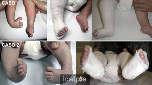
Also called clubfoot. It is an abnormal position of the foot that occurs from birth. Babies are born with their feet turned inward and downward. It requires treatment with corrective casts, followed by Achilles tendon tenotomy, and then maintaining the achieved correction using nighttime orthotics. All these therapeutic procedures are part of the Ponseti method.
It is a foot that presents a flattened arch, meaning the foot’s bridge is reduced. The patient may experience limited mobility, and it can be painful.
It is a foot with an excessive plantar arch or bridge. They may present pain and plantar calluses.
The foot rests on the inner part of the heel, and the ankle tilts inward.
The heel and toes rest on the outer part of the foot.
The patient only places the tips of their toes down and does not let the heel touch the ground.
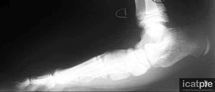
Radiological image of a patient with flat and equinus foot secondary to a neurological disease.
It is defined as a dysfunction of the hip joint that leads to improper functioning. This pathology usually presents from birth, although some cases are diagnosed during childhood. Most cases are mild and resolve spontaneously or with the use of orthotics. However, there are cases of congenital hip dislocation that require surgery.
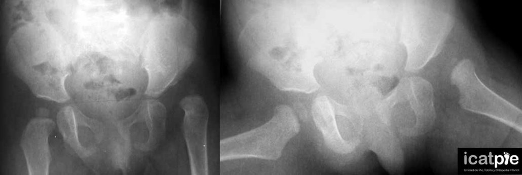
Perthes disease (Legg–Calvé–Perthes) is a condition of the hip in children between the ages of 2 and 12, with a peak incidence between 4 and 8 years old. It involves necrosis of the femoral head to varying extents. The disease progresses from necrosis to the complete regeneration of the femoral head, a process that takes several years during the child’s growth. The patient typically reports pain and limited mobility in the affected hip. Treatment ranges from sports restriction and physiotherapy to surgery. Deciding on the appropriate treatment should be individualized based on the patient’s age and the extent of the damage. Therefore, it is important for the child to be evaluated by a pediatric orthopedic specialist to determine the most appropriate treatment.

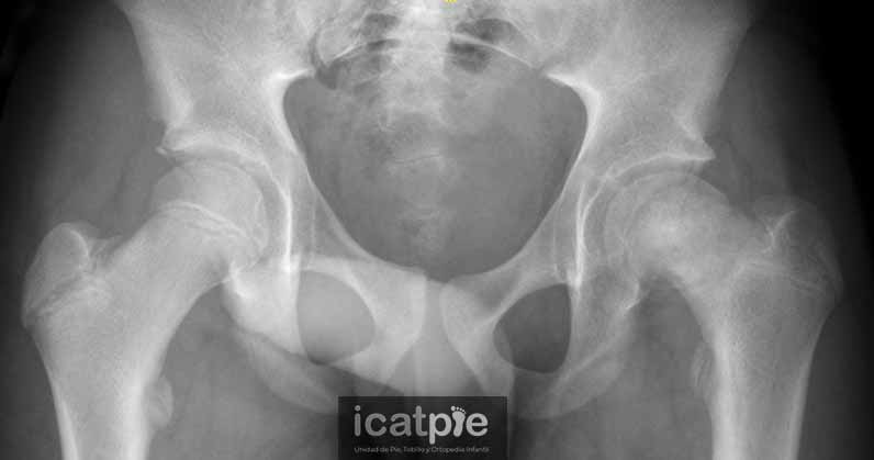
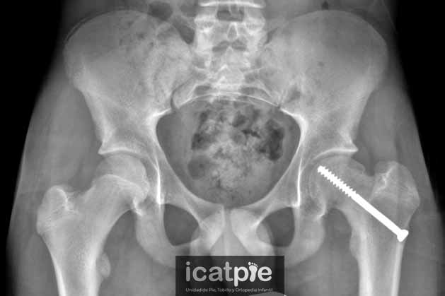
Slipped capital femoral epiphysis (SCFE) is a condition that typically affects pre-adolescents (around 12 years old in girls and 13 years old in boys). It involves the displacement of the femoral head over the femoral neck, as if a fracture occurs in the femoral head without any trauma. The patient usually presents to the emergency department with more or less severe pain in the hip and limited mobility. This condition requires urgent surgical treatment to fix the femoral head to the rest of the bone to prevent further displacement.