Unit of Foot and Ankle
Specialists in the study, diagnosis, and application of surgical techniques for foot surgery.
Inicio › Medical units › Unit of Foot and Ankle › Fractures of the Foot and Ankle
Fractures of the Foot and Ankle
When we talk about fractures of the foot and ankle, we refer to any type of break located in the bones that form these joints. In the case of the ankle, we refer to fractures of the tibia and fibula, and in the case of the foot, fractures of the tarsal bones (talus, calcaneus, navicular, cuboid, or cuneiform), metatarsals, and phalanges.
The foot is a crucial element in locomotion and supports a biomechanical load of up to seven times the body’s weight. From this, we can deduce that these types of fractures will be very common, and therefore, it is essential to make an early diagnosis and provide appropriate treatment.
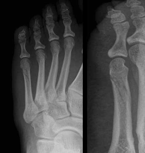
(*In the image, we can see X-rays of an ankle fracture)
What causes fractures in the foot and ankle?
Fractures occur due to trauma to the bones, with the most common causes being:
- Tripping or falling.
- Impacts from heavy objects, a situation frequently seen in both the workplace and at home.
- Traffic accidents.
- Sports accidents.
- Having conditions that lead to decreased bone density (osteoporosis), which makes bones more fragile in the event of falls.
- Advanced age or suffering from conditions that make walking difficult or impair balance, such as certain neurological or rheumatic diseases.
- Poor lighting at home.
- Improper vehicle driving.
- Working in environments that involve a risk of falling from a height or being impacted by heavy objects.
- Engaging in high-impact sports.
- Playing sports with improper equipment or technique.
What symptoms may occur?
The most common symptoms typically include:
- Pain, swelling, and immediate tenderness in the area of the fracture
- Deformity of the affected area
- Bruising
- Difficulty or inability to walk or stand
How is it diagnosed?
The first step is to take a detailed medical history of the patient to understand any pre-existing conditions and the mechanism of the injury.
Next, a thorough physical examination is performed. During the examination, it is common for the patient to show hypersensitivity and pain when the affected area is palpated.
Then, an X-ray of the foot or ankle is taken to confirm the suspicion of a fracture and identify which bones have been affected. In cases where the X-ray does not provide enough information to make an accurate therapeutic decision, a computed tomography (CT scan) may be performed.
What treatment is advised?
The treatment will depend on the bone fractured and the severity of the injury.
There are three types of treatment:

Immobilization without reduction is the treatment for milder fractures, where the bone fragments have not been displaced upon breaking.
In these cases, we proceed to immobilize the bone so that the ends can heal correctly. A cast is usually used, although depending on the bone fractured, other immobilization devices such as orthopedic boots, rigid sole footwear, or in the case of phalanges, bandaging with tape and gauze may be used.
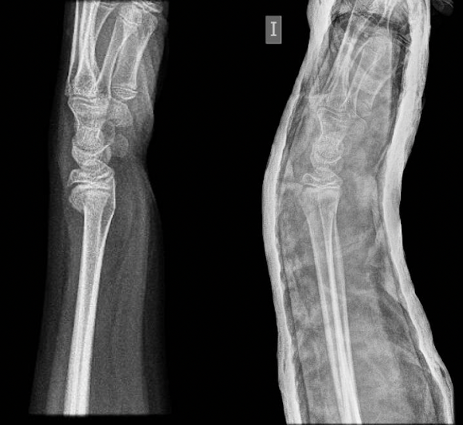
We refer to reduction as the process of manipulation by the specialist doctor of the fractured bone fragments to return them to their correct position. This technique is used when dealing with displaced fractures, meaning the bone fragments are not aligned. Depending on the severity, anesthesia or muscle relaxants may be used before performing the procedure. Once the bone fragments are in the correct position, a cast is applied to maintain the reduction until the fracture heals (or consolidates).

Radius fracture in a pediatric patient. The fracture had displacement, which required reduction and the application of a cast to maintain the reduction until healing.
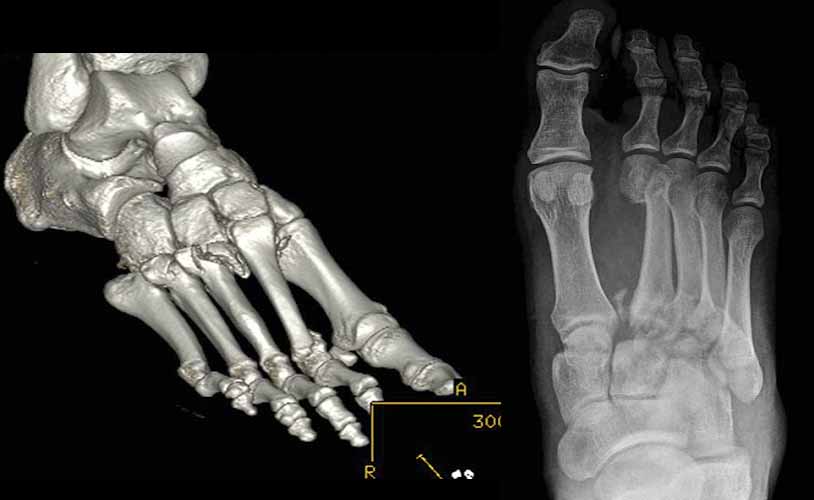
In cases where fracture reduction is difficult to achieve using a cast, the specialist will recommend reducing and maintaining the alignment of the bone fragments using metal implants such as plates, pins, screws, or nails. Once the fracture has healed, if the implants cause discomfort or skin irritation, they may be removed.

Conventional X-ray image (right) and CT scan (left) of fractures affecting the so-called Lisfranc joint, which includes the midfoot joints between the cuneiforms and metatarsals. These are displaced fractures that will require surgery.
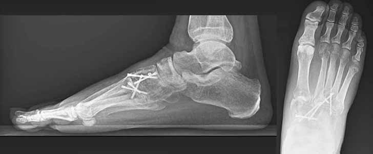
Postoperative X-rays of the previous patient. Through open surgery, the fractures were reduced, and screws were implanted to maintain the reduction until healing.